In the spring of 1972 (my second year in college) I worked in the Department de Pathologie at the University of Geneva, Switzerland. There I learned a variety of lab techniques (I had previously worked in a different lab, doing various jobs, at Harvard Medical School), including tissue culture and time-lapse photographic microscopy.
The director of the lab, Guido Majno (a close personal friend of my father's), had become interested in the phenomenon that wounds heal much more slowly when circular than when straight, or rectangular. He called this the "turtle" effect because he imagined a circle of turtles walking toward each other and getting stuck when their shells collided, leaving a circular gap (analagous to slow healing).
I decided to run an experiment on this by growing fibroblasts in culture, and creating small wounds in the cultured layer, then creating a movie of them as they grew in. (I did this surreptitiously, after hours, because I knew I would not have been given permission to do this on my own.) The results were astonishing: in time-lapse we could see the cells slowly moving toward each other, then one would "extend an arm" (cytoplasm) toward another, they would wrap each other and pull, and then suddenly hundreds of cells would pile on top of each other. In other words, we could see mechanistic aspects of wound healing in vitro.
My report on the turtle experiments may be found here.
Guido raised his eyebrow when I told him about what I had done (right at the end of my stay in his lab), but when he watched the movie he also thought it was amazing. He promised to follow up, repeat, and publish the results. This never happened. My impression is that this was about the time that people were starting to work out mechanisms of fibroblast locomotion (e.g., Theriot, JA and Mitchison, TJ (1992) J. Cell Biol., 118: 367-377, and refs therein).
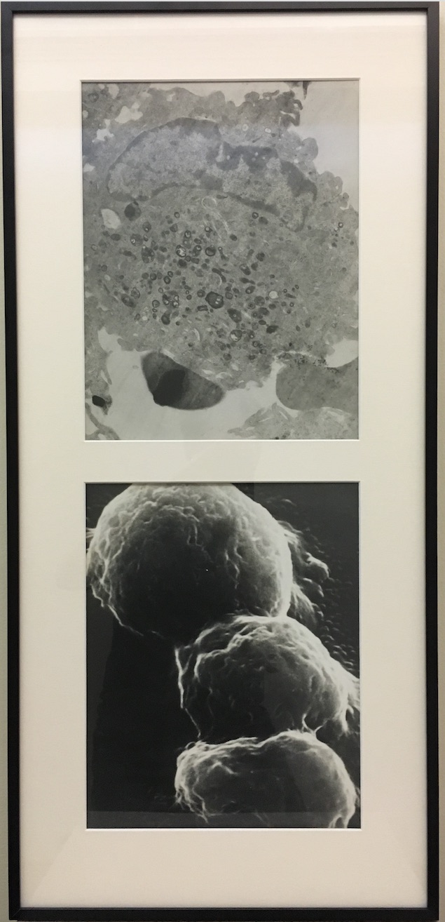
In my earlier jobs (especially in high school and just after) I had learned electron microscopy. I saved an electron micrograph of an alveolar macrophage, because I had done all the work, from harvesting the cells from the animal, to embedding the tissue in epoxy, to slicing with a diamond knife, to running the microscope, and finally developing and printing the picture. It is the top one here: